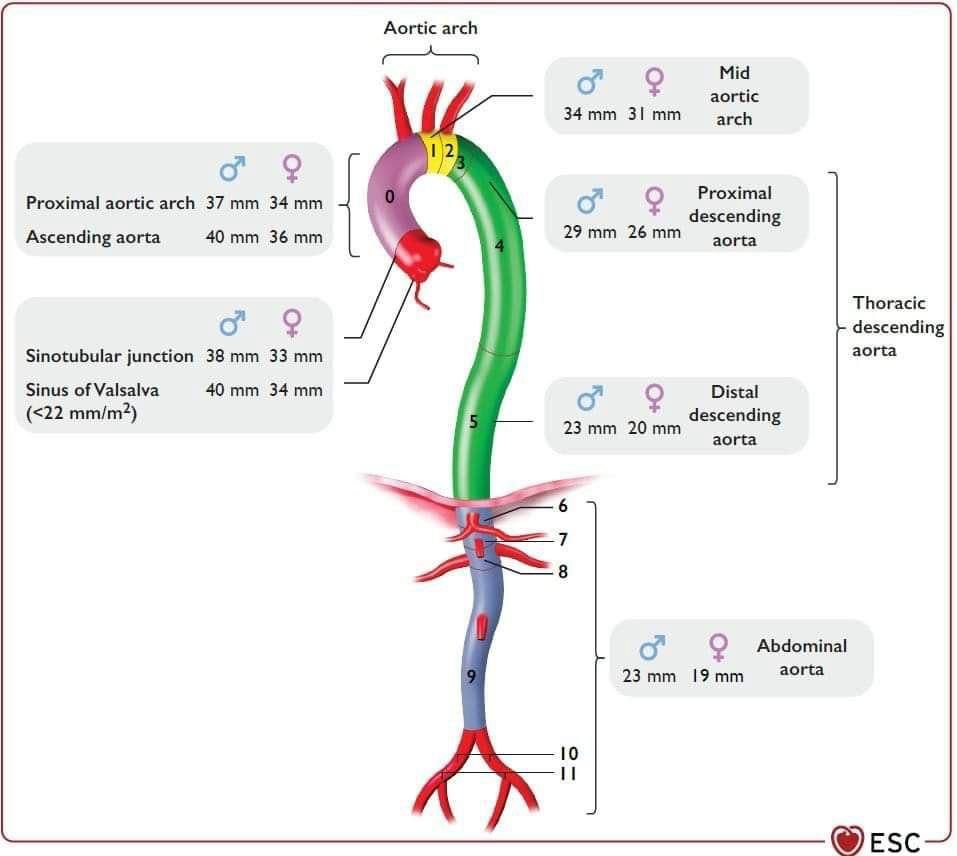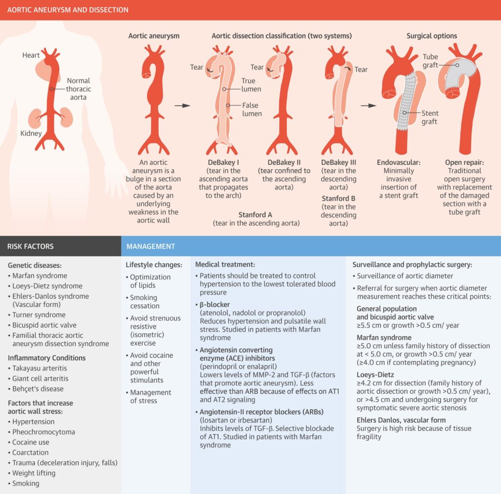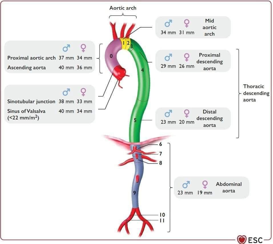
Aortic aneurysm and dissection
An aneurysm is a permanent, localized arterial dilation to more than 50% of the normal diameter. The histopathology of some aortic aneurysms involves medial degeneration (formerly called cystic medial necrosis), characterized by elastic fiber loss from the medial layer, loss of vascular smooth muscle cells, and proteoglycan deposition. Altered microribonucleic acid expression is implicated in increased activity of matrix metalloproteinase (MMP)-2 and -9 , which lyse elastic fibers and break down the extracellular matrix. The media within the aneurysm is also remodeled, with smooth muscle cells oriented randomly, instead of in organized layers parallel to the aortic lumen. Transforming growth factor (TGF)-β, a regulatory cytokine expressed by arterial wall vascular smooth muscle cells, modulates MMP activity. In a mouse aortic aneurysm model, disrupting TGF-β signaling prevented aneurysm formation .There are 2 TGF-β signaling pathways, and both play roles in aneurysm formation . In the canonical pathway, TGF-β binds at the cell surface to its type I or type II receptors. The complex phosphorylates Smad2 or Smad3, resulting in Smad4 recruitment and nuclear translocation, and Smad-dependent gene transcription. In the noncanonical pathway, TGF-β phosphorylates extracellular signal-regulated kinase (ERK), Jun N-terminal kinase, and p38.